|
A veces observamos pequeños organismos cuando miramos muy cerca nuestro acuario, microbios, gusanos, etc. y algunos que haría falta una lupa o un microscopio para poder verlos. . . . . .
. . . . podríamos clasificar los organismos que pululan en el agua de nuestro acuario por su tamaño:
1 - Hasta 2 milimetros mesoplancton
2 - De 2 a 10 milimetros macroplancton
3 - Más de 10 milimetros megaloplancton
Entre 0 y 2 milimetros necesitariamos un microscopio para poder observarlos, algunos infusorios como los paramecios casi se pueden observar a simple vista o con una lupa.
 Esta foto pertenece a: Nino Santamaria
El plancton se divide en:
HOLOPLACTON: es el plancton permanente, que a lo largo de todas sus etapas conserva un tamaño microscopico.
MEROPLACTON: es una etapa primaria en el desarrollo de algunas espacies, huevos y larvas de insectos, moluscos, anfibios, peces, etc.
Otra clasificación es el plancton vegetal (fitoplancton) y el plancton animal ( zooplancton)

Fotos Cristina Eisman - Pertenece a: regmurcia.com
Un acuario nuevo al principio esta carente de materia orgánica en disolución, pero con el tiempo y dependiendo de la temperatura, luz, y nutrientes ira floreciendo una colonia de seres vivos, normal y beneficiosa en el pequeño ecosistema que hemos creado, los rayos UVA, (lámparas germicidas), el cloro ó el ozono, y productos químicos como el permanganato de potasio reducen drásticamente la colonia beneficiosa de seres vivos microscópicos, por lo que utilizaremos estos medios de esterilización muy cuidadosamente o simplemente no los utilizaremos.
Algunos organismos los veremos porque están dentro del tamaño de macroplancton ó megaloplancton y los microscópicos no los veremos a simple vista, la mayoría de ellos son inocuos, e incluso beneficiosos para nuestro acuario, realizan funciones de filtración y clarificando el agua reduciendo elementos orgánicos que hay en disolución o en suspensión.
¿Como llegan estos organismos a nuestro acuario?
Pueden llegar a través del agua del grifo, sobre todo en zonas que no está muy tratada el agua potable, pero también pueden llegar en forma de esporas por el aire, los organismos enquistados y los huevos pueden resistir la congelación ó productos químicos desinfectantes muy fuertes, también a través de plantas importadas, en la piel o en el interior de los peces ó las heces de algún pez, de alguna forma el acuario es un ciclo cerrado agua y tarde o temprano pulularan muchos tipos de microbios, afortunadamente gracias a ello se completará el ciclo del nitrógeno y la vida no se acabará.
Habrá que distinguir de entre estos organismos los que son beneficiosos, y los que puedan causar enfermedades o parasitar a los peces ó plantas, gusanos de la piel, branquias, ácaros, hongos, bacterias, algas, etc. mientras los peces no den síntomas de malestar, irritaciones o movimientos convulsivos, pensaremos que esos bichitos hacen una función intermedia en la cadena de alimentación.
Prestaremos especial atención a las especies capturadas en la naturaleza sean peces o plantas, ó alimento vivo, tubifex, larvas de mosquito, dafnias, cyclops, etc. desechando lo que tengamos duda y sometiendo a observación, las plantas se deben desinfectar siempre, ó dejarlas un par de días en un acuario vacío de peces.
Los gusanillos que se desplazan por los vidrios son detritívoros, miden desde unos pocos milímetros a más de un cm. y finos como un hilo de coser, son inofensivos pero si tenemos demasiados es por el exceso de comida sobrante, algunos anabantidos los devoran, pueden ser planarias si tienen la cabeza en forma de flecha y estan sobre las plantas y cristales, y los alevines muy pequeños pueden ser atacados en su fase larvaria, para evitar su proliferación es conveniente hacer sifonados de grava de vez en cuando.

Esta foto pertenece a: ZOOINVERTEBRADOS
Muchos de los organismos microscópicos que no vemos, los infusorios por ejemplo que miden de 0.5 a 2 milímetros, de las que hay centenares de especies, paramecios, vorticelas, etc. son muy beneficios porque filtran el agua y sirven de alimento para los alevines más pequeños.
Las Hydras, son parecidas a las anemonas de mar pero mucho más pequeñas y solo peligrosas para los alevines recién nacidos, normalmente llegaran a nuestro acuario en alguna planta.

Esta foto pertenece a: microscopy-uk.org.uk
MICROCRUSTACEOS, hay más de 7000 especies de hasta un milímetro ó dos de longitud se alimentan de detritus, aumentados con un microscopio se parecen a artemias o gambas y son un excelente alimento para los alevines después de haber pasado la etapa de los infusorios, los alevines de discos se los pueden comer desde el principio.

Esta foto pertenece a: Invertebrados de agua dulce
Por último las microalgas flotantes de tono verde son el principal alimento del zooplancton, larvas de mosquito, gusanos, amebas, paramecios, etc.

Esta foto pertenece a: aquaticscape.com
|
|
|
Fuente : El acuario y la electronica
Una guía sencilla para pequeña y microscópica vida de la charca con enlaces a recursos Micscape
Uno de los temas más gratificantes para el estudio con el microscopio son organismos de agua dulce. Métodos de recolección simples incluyen apretando las plantas de agua en una jarra y para especies libres de natación, una multa-Meshe
La forma más fácil de recoger los microorganismos y otra pequeña vida de la charca es exprimir el agua de las plantas de agua o espuma de la charca en un recipiente.

Otro método consiste en raspar el crecimiento de las plantas de agua u otras cosas que están cubiertos por un crecimiento verde o marrón . Una tarjeta de crédito de edad hará un excelente raspador pero tenga cuidado de no dejarlo caer en el estanque !

Se recomienda una red de plancton de las especies de natación libre ( planctónicas ) . Esta es una red hecha de una tela de malla muy fina con un pequeño recipiente en el extremo. También puede utilizar la red de plancton para concentrar el material expulsado de plantas de agua .
Las páginas de tablas y enlaces son una guía para algunos grupos comunes de organismos de agua dulce más pequeños (microscópicos de unos pocos milímetros de tamaño). Si no está familiarizado con un organismo, ver qué dibujo y características
El principiante también disfrutan de explorar la inmersión estanque virtual; haga clic en las criaturas en el tarro de aprender acerca de algunos de los organismos de agua dulce plebeyo.
Bacteri
unicelular , puntos o filamentos , apenas visible con un aumento más fuerte , las cianobacterias son más grandes
Introducción a las bacterias
Spirochaetes
unicelular, con diminutos pelos o pseudópodos
Ir a la visión general protozoos: por ejemplo, ciliados, ameba, heliozoos, euglenoids
|
~ Pond Life Identification Kit ~
|
|
A simple guide to small and microscopic pond life
with links to Micscape resources
|
|
One of the most rewarding subjects for study with a microscope are freshwater organisms. Simple collecting methods include squeezing water plants into a jar and for free swimming species, a fine-meshed plankton net is recommended. For simple tips see how to collect microscopic pond life.
The table and linked pages are a guide to some common groups of smaller freshwater organisms (microscopic to a few millimetres in size). If not familiar with an organism, see what drawing and features it most closely resembles in the table and then follow the links.
The beginner may also like to explore the virtual pond dip; click on the creatures in the jar to learn about some of the commoner freshwater organisms.
|
|
|
Bacteria
|

|
single celled, dots or strands, just visible with strongest magnification, cyanobacteria are larger
|
Introduction to bacteria
Spirochaetes
|
|
Protozoa
|

|
single celled, with tiny hairs or pseudopodia
|
|
|
Algae
|

|
single celled, mostly green, sometimes yellow-brown
|
|
|
Rotifers
|

|
wheel-like, hairy appendages, transparent, free swimming or attached 0.2 - 1 mm
|
'Smallest page on the web' - rotifers
|
|
Gastrotrichs
|

|
two tails, hairy, round mouth opening
0.1 - 0.5 mm
|
No Micscape resources. (Articles welcomed!)
|
|
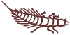
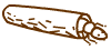
Worms
|

|
long thin body, many non related forms
|
|
|
Bryozoa
|

|
plant-like or jelly-like colony, crown of tentacles
individuals: 0.25 - 5 mm
|
Bryozoans
Pond fairies - Plumatella repens
|
|
Hydra
|

|
green brown or colourless, body and tentacles contract and stretch
extended: 20 mm
|
Introduction to hydra
Hydra in 3D
Hydra oligactis
Video clips of a hydra
|
|
Water bears
(Tardigrades)
|

|
8 stumpy legs, slow moving
<1 mm
See gallery links on the right for some of the finest video clips on the Web of these cute critters!
|
Hunting for 'bears' in the backyard
The incredible water bear
Water bear video gallery I
Water bear video gallery II
|
|
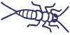
Arthropods
|


|
jointed limbs; many groups e.g. crustaceans ('water fleas'), mites
|
|
|
other Arthropods:
Insect stages
|

|
wide variety of forms
|
|
|
Protozoa~ Some common freshwater types with links to Micscape resources
|
Protozoa are a very diverse group of organisms that vary widely in size, shape, features and habit. This page gives an overview of some commonly found freshwater protozoa.
The protozoa have been grouped by their major features. Some of these are artificial groups (i.e. not necessarily related to their taxonomy) but are convenient ones for the pond dipper. More about the classification of algae and protozoa. |
| Group |
|
Key features
|
Micscape links
|
|
Flagellates
(those that photosynthesise are often classed as algae)
|

|
one or more flagella (whip-like cilia), phytoflagellates are green/ photosynthesise, zooflagellates are not green
<0.4 mm
|
Flagellated protozoa - includes Euglena, Volvox
Some other common types: Monads e.g. Bodo, Choanoflagellates (flask-shaped with flared collar)
|
|
Amoeba
|

|
move with pseudopods
0.02 - 5 mm
|
Amoeba - Protozoa portraits
Amoeba - Video gallery
Amoeba - Smallest page on the web
Amoeba proteus - imaged by different illumination methods
|
|
Shelled amoeba
|

|
amoeba with a shell e.g. of sand grains
0.1 - 0.4 mm
|
Protozoan houses - testate amoeba, Arcella, Nebela
Some other common types: Chaos, Pelomyxa
|
|
Heliozoans
'Sun animalcules'
|

|
immobile, spherical with radiating hair-like pseudopods
0.01 - 1 mm
|
Smallest page on the web - Heliozoans, Actinosphaerium
Some other common types: Actinophrys, Acanthocystis
|
|
Ciliates - Peritrichs
|

|
cylindrical or bell-shaped bodies, undulating membrane of cilia, some stalked, often colonial and attached to animals or plants bell: <0.25mm
|
Bell animalcules in 3D - Campanella
Ophrydium - colonial, unstalked
Vorticella - observations on its motile stage
Some other common types: Vorticella, Carchesium
|
|
Ciliates - Suctoria
|

|
on water plants and other animals, adult ciliates have lost cilia, sticky tentacles capture prey <0.7 mm
|
Acineta - Suctoria, ciliates in disguise
Podophyra - an interesting ciliate
Some other common types: Tokophyra, Dendrocometes (lives on gill plates of f/water shrimp)
|
|
Other ciliates
|

Coleps

Lacrymaria

Paramecium

Stentor

Spirostomum
|
various, mostly free living forms
cell usually of a fixed shape but can be contractile, or extending neck, cilia of various forms, fixed mouth
0.01 - 4 mm
|
Smallest page on the web - ciliates, Euplotes, Stylonichia, etc.
Actinobolina vorax
Amphileptus
Coleps - a voracious protozoa
Coleps & Urotricha - predation on rotiferColpidium -
Protozoa portraits
Colpoda - emerging from resting stage
Didinium - a master feeder
Dileptus - a carnivorous protozoa eating Litonotus
Dileptus - notes on it eating Cyclidium
Lacrymaria olor - the 'giraffe' of the protozoan world
Lacrymaria olor - microscopic 'Loch Ness monster'
Litonotus - being eaten by Dileptus
Paramecium - an introduction
Paramecium - Protozoa portraits
Paramecium - by phase contrast
How to study a 'pair of mecia' parts I-III- extensive articles on microscopy techniques to study paramecium's features. Ideal for students.
Stentor - introduction
Stentor - in 3D
Spirostomum- one of the largest ciliates
Spirostomum - observations on its feeding habits
Tillina
Features of ciliates:
Video gallery - beating cilia
Cilia - an overview
|
|
Algae ~ Some common freshwater types with links to Micscape resources
|
Algae are a very diverse group of organisms that vary widely in size, shape, colour and habit. Ten or so phyla are represented in freshwater. This page gives an overview of some commonly found freshwater algae.
The algae have been grouped by their major features. Some of these are artificial groups (i.e. not necessarily related to their taxonomy) but are convenient ones for the pond dipper. More about the classification of algae and protozoa. |
|
Group
|
|
Key features
|
Micscape links
|
|
Flagellated forms (some are also often classed as protozoa)
note: These flagella are hardly visible, only with strong magnification.
|
|
|
Euglenoids
|

|
green, flagella (whip-like cilia), free-swimming, red eye spot, body is flexible <0.4 mm
|
Flagellated algae/protozoa - includes Euglenaa heart shaped euglenoid: Phacus
|
|
Dinoflagellates
|

|
brown, 2 flagella, (1 in girdle), free-swimming, tough armour <0.4 mm
|
Peridinium - Stars of the Marshes
|
|
Green algae
(Chlorophyta)
|

|
spherical colonies, cells with 2 flagella
Volvox: 0.5 - 2mm
|
Volvox - the jewel of the pond
Volvox in 3D
Some other smaller colonial flagellates: Gonium, Eudorina, Pandorina,Sorastrum (rare), Synura, Uroglena
|
|
not all green algae are green
|

|
tiny, green/red, often in bird baths
<0.05 mm
|
Haematococcus
Haematococcus - image
another small flagellated green algae: Chlamydomonas
|
| There are many other small flagellated algae of non related groups. Classification is difficult. |
|
Non-flagellated forms
|
|
|
Blue-green algae(cyanobacteria)
|

|
blue-green, often slow locomotion,
used to be considered algae but more related to bacteria cells<0.05 mm colonies can be many mm
|
Smallest page on the web - 'bacteria' - image of a cyanobacteria
|
|
Diatoms
|

|
usually brownish, silica cell wall in two parts, solitary or colonial, some have a slow gliding motion
<0.5 mm
|
Smallest page on the web - 'diatoms'
Those who live in glass houses'
Diatoms on strings
Bacillaria - 'Carpenter's rule' diatom
Library search - type diatom for articles on f/water & marine species
Test diatoms - species to test optics
|
|
Desmids
(Gamophyta: conjugating green algae)
|

|
green, no flagella, mainly solitary, some colonial, various shapes, two semi-cells which are mirror images
<0.5 mm
|
What are desmids?
Smallest page on the web - desmids
Bill Ells' page - extensive articles on various species and aspects of desmids
Desmids in 3D
Desmid gallery
|
|
Green algae
(Chlorophyta)
|

|
green, don't move, no flagella, not attached to a surface
starshaped colony: Pediastrum <0.3 mm
bottom right: Scenedesmus <0.03 mm
|
Pediastrum - image only
Pediastrum - the 'star' of the pond
Asterococcus
Some other common 'non flagellates': Chlorella (symbiont in several organisms), Scenedesmus, Dictyosphaerium
|
|
Other algae of various growth forms
|
|
|
Water net
|

|
a sock-like colony, green algae (related to Pediastrum) up to 20 centimeters
|
Hydrodictyon - the water net
|
| Some other common algae of various types: Botrydium, Chaetophora, Coelochaete, Enteromorpha |
|
Filamentous forms
|
|
|
Pond scum
(Gamophyta: conjugating green algae)
|

|
non-branching, green, chains of cells with distinctly shaped cell contents
cell with <0.1 mm. length: centimeters
|
Spirogyra - chloroplasts like helix
Spirogyra in 3D
Zygnema - starshaped chloroplasts
Mougeotia has plate-like chloroplasts
|
|
Other non-branchingforms
|

|
several non related groups
|
Oedogonium
Some other common filamentous types: Tribonema, Ulothrix, Vaucheria
|
|
Branching forms
|

|
mostly green algae
|
Cladophora, Draparnaldia, Stigeoclonium
|
|
Red algae(Rhodophyta)
|

|
mainly marine, but some freshwater forms, not always red
|
Batrachospermum
Porphyridium
|
|
Worms ~ Some common freshwater types with links to Micscape resources
|
There are a number of groups of worms that occur in freshwater. Many aren't microscopic but are shown here to give an overview of these groups. Some common organisms that may be confused for a worm are also included.
Note: this table isn't comprehensive, there are several other freshwater groups like ribbonworms and horsehair worms. |
|
|
|
Key features
|
Micscape links
|
Flatworms
(Platyhelminthes)
|
 |
flattened, 2 or more eye spots, move in gliding fashion
1 - 15+ mm
|
image of flatworm
|
Segmented worms
(Annelids)
|

 |
oligochaetes: 1.5 mm. to >2cm long, hair bundles, (includes earthworm andTubifex)
leeches: characteristic 'leech-like' motion, suckers each end >1 cm
|
image of oligochaete
|
|
|
 |
move very frantically, often in 'S' curves,
0.2 - 10 mm
|
image of nematode
|
not to be mistaken for worms:
|
|
Hydra, has tentacles
extended: 2 cm
|
Introduction to hydra
|
|
larger ciliated protozoa like Spirostomum (has very fast contraction)
|
Spirostomum
|
 |
insect stages like mosquito larva, these have a more distinct head than the worms above, also antennae/legs
|
Go to arthropods overview
|
|
|
Arthropods~ Some smaller freshwater types with links to Micscape resources
|
Arthropods are characterised by jointed limbs and include major groups like the crustacea, insects, spiders and mites. They have many segments, a tough outer skeleton and many modified limbs. There are many microscopic and macroscopic types that occur in freshwater.
Notes:
There are several other groups with limb-like structures that aren't arthropods. Check rotifers and hydra on the first page. |
This page with links hopefully gives a useful overview, but it's neither a formal identification guide nor comprehensive. Water bears have been included here as well as on the first page, because some resources regard them as an arthropod phyla.
Acknowledgements: Many thanks to all the Micscape contributors whose articles this guide links to. For clarity their names are omitted in the links above.
An Introduction to Microscopy
|
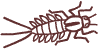
Insect stages ~ Some larvae, nymphs and adult insects that live in freshwater
|
In pond water you will come across many insects, often perfectly adapted to the aquatic environment. Some species are entirely aquatic, whereas other insects only live in the water during their larval stages or as nymphs. When insects undergo a metamorphosis we call the immature form larva. When they gradually transform via moults into the adult form the young stages are called nymphs. This pages gives a simple overview of these stages and some of the adult forms. There are no Micscape links yet, articles welcomed!
Note on size: Many aquatic insects and their immature stages can vary in size from a few mm to 3cm or more depending on e.g. maturity and species, so sizes have been omitted for most groups. The shape and general features are a more reliable guide to a group than size. |
|
Group
|
|
key features
|
|
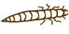
Alderfly nymph
|
|
one tail, long filaments along the abdomen
|
|
Caddisfly larva
|
|
most species build a cylindrical case for protection, each species makes a distinct case from different material
|
|
Stonefly nymph
|
|
two jointed tails
|
|
Mayfly nymph
|
|
three jointed tails, leaf-like (or other shaped) 'gills' on its sides
|
|
Damselfly nymph
|
|
three leaf-like tail appendages (gills), bizarre extendable jaws
|
|
Dragonfly nymph
|
|
robust, no tail appendages, bizarre extendable jaws
|
|
Water bug
nymph/adult
|
|
no jaws, like all water bugs they possess a tube-like beak, the nymphs don't have wings,
Some common forms: Backswimmer, water boatman. On the water surface: Pond skater
|
|
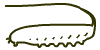
Water-beetle larva
|
|
strong jaws, long segmented body, short legs
|
|
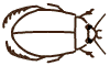
Water beetle adult
|
|
strong jaws, tough shield, many water beetles are fierce predators
|
|
Springtail
|

|
the grey spring-tail (the most primitive insect group) Podura aquatica lives on the surface of the water, often in large numbers, 0.5-2.5mm
Visit the Postal Microscopical Society (UK) Springtail Group site for overview and projects
|
|
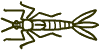
Mosquito larva
|
|
with a long slender body, often moves in S-shaped curves,
|
|
Dronefly larva
|
|
this so called rat-tailed maggot has a long tubed tail for breathing
|
|
Other Arthropodsthat are not insects
|
|
|
This page with links hopefully gives a useful overview, but it's neither a formal identification guide nor comprehensive.
An Introduction to Microscopy
Comments to the compilers Wim van Egmond and Dave Walker are welcomed.
Microscopy UK Front Page
Micscape Magazine
Article Library
All images © Wim van Egmond
© Onview.net Ltd, Microscopy-UK, and all contributors 1995 onwards. All rights reserved. Main site is at www.microscopy-uk.org.uk with full mirror at www.microscopy-uk.net.
|
|
|